Voiding Cystourethrogram (Pediatric)
Voiding Cystourethrogram (Pediatric)
What is a Voiding Cystourethrogram?
During a voiding cystourethrogram, the bladder is filled with contrast material to see if any reverse flow (called reflux) occurs from the bladder into the ureters (the drainage tubes from the kidneys). Checking for reversed flow is important because the flow should be one way only: down from the kidneys, through the ureters and into the bladder. Reflux, often associated with infections, can damage the kidneys and cause scarring and poor function years later. In addition, the VCUG will check the ability of the child’s bladder to empty properly and will also show any abnormalities of the urethra (the final channel from the bladder to the skin surface).
How is a VCUG performed?
A VCUG is commonly performed after the first urinary tract infection in children. Urinary tract infections are much more common in girls because they have a short urethra, which allows the bladder to be more easily infected from the outside.
- For a child under the age of 3, we may use a special device called an octagon board where the child is secured during the exam like a little papoose. This does not hurt the child, but is done to help reduce the amount of radiation the child receives during the exam by restricting unwanted motion. It also allows the Radiologist to turn the child quickly onto their side so as not to miss any crucial anatomy during the exam.
- On a child over the age of 3 years of age, they will lay on towels on the x-ray table with one parent remaining with the child.
A preliminary x-ray is taken of the abdomen. To catheterize the bladder, a specially trained technologist will first cleanse the area around the urethra with an antiseptic solution before slipping the small, soft catheter into the urethra. A small piece of tape will secure the catheter so it will not slip out. The child might experience the sensation that they have to go to the bathroom but will be voiding through the catheter.
The contrast (iodine mixed with sterile water), will be hung like an I.V. bottle and will be connected to the catheter. The contrast is warm and most children cannot feel the bladder filling. As the contrast flows into the bladder by a gravity drip the Radiologist will be taking x-rays of the kidneys, ureters, bladder and the urethra as the child voids. The child will experience some discomfort when the bladder fills to capacity but this will be relieved when the child voids. The contrast stops dripping when the child’s bladder is full. The child will be asked to void on the table in towels/cloth diapers. An older male child may use a urinal. It is important for the child to void on the table so the Radiologist can identify reflux during the voiding cycle. Sedation is not recommended because it can interfere with the child’s ability to void.
Doctors

David Alan Bloom, MD
Professor
Urology, Pediatric Urology, Surgery

Michael Anthony Di Pietro, MD
Professor Emeritus
Pediatric Radiology, Pediatrics, Diagnostic Radiology

Kathleen M S Gebarski, MD
Clinical Assistant Professor
Pediatric Radiology, Diagnostic Radiology

Ramiro Hernandez, MD
Professor Emeritus
Pediatric Radiology, Diagnostic Radiology

Anastasia Louise Hryhorczuk, MD
Clinical Associate Professor
Diagnostic Radiology, Pediatric Radiology

Aparna Joshi, MD
Clinical Associate Professor
Diagnostic Radiology, Pediatric Radiology

Maria Fernanda Ladino Torres, MD
Clinical Associate Professor
Pediatric Radiology, Diagnostic Radiology

Gary Dean Luker, MD
Professor
Diagnostic Radiology, Pediatric Radiology

Swati Shah Mody, MBBS
Clinical Associate Professor
Diagnostic Radiology, Pediatric Radiology

Peter Jackson Strouse, MD
Professor
Diagnostic Radiology, Pediatric Radiology

News & Stories
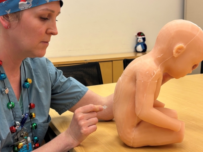
From simulation to practice for doctors
New mouse model for liposarcoma can help uncover new therapies
Rare infant triplet overcomes one-in-a-million type of liver cancer

An AI model that can read and diagnose a brain MRI in seconds
Hospital partnership improves follow up scans, decreases long term risk after aortic repair