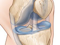
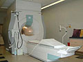
Magnetic resonance imaging (MRI) is a test done with a large machine that uses a magnetic field and pulses of radio wave energy to make pictures of the knee, Opens dialog. Muscles, ligaments, cartilage, and other joint structures are often best seen with an MRI. In many cases an MRI gives information about structures in the body that cannot be seen as well with an X-ray, ultrasound, or CT scan.
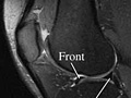
For an MRI test, you are placed inside the magnet so that your knee is inside the strong magnetic field. An MRI can find changes in the structure of organs or other tissues. It also can find tissue damage or disease, such as infection or a tumor. Pictures from an MRI scan are digital images that can be saved and stored on a computer for further study. The images also can be reviewed remotely, such as in a clinic or an operating room. Photographs or films of selected pictures can also be made.
In some cases, a contrast material may be used during the MRI scan to show certain structures more clearly in the pictures. The contrast material may be used to check blood flow, find some types of tumors, and show areas of inflammation or infection. The contrast material may be put in a vein (I.V.) in your arm or directly into your knee.
Magnetic resonance imaging (MRI) of the knee is done to:
Check for the cause of unexplained knee pain or the knee giving out for no reason.
Find problems in the knee joint, such as arthritis, bone tumors, or infection, or damaged cartilage, meniscus, Opens dialog, ligaments, or tendons.
Find out if a knee arthroscopy is needed.
MRI may also find a bone fracture when X-rays and other tests do not give a clear answer. MRI is done more commonly than other tests to check for certain bone and joint problems.
In general, there's nothing you have to do before this test, unless your doctor tells you to.
Tell your doctor if you get nervous in tight spaces. You may get a medicine to help you relax. If you think you'll get this medicine, be sure you have someone to take you home.
Information about Magnetic Resonance Imaging (MRI) of the Knee
You will need to remove all metal objects (such as hearing aids, dentures, jewelry, watches, and hairpins) from your body. These objects may be attracted to the powerful magnet used for the test.
You will need to take off all or most of your clothes, depending on which area is examined. (You may be allowed to keep on your underwear if it's not in the way.) You will be given a gown to use during the test. If you are allowed to keep some of your clothes on, make sure your pockets are empty.
If you wear a medicine patch, you may need to remove it. The MRI can cause burns with some patches.
During the test
During the test, you will lie on a table that is part of the MRI scanner. The table will slide into the space that contains the magnet. A device called a coil may be placed over or wrapped around the area to be scanned.
Some people feel nervous inside the MRI scanner, Opens dialog. If feeling nervous keeps you from lying still, you can be given a medicine (sedative) to help you relax.
Inside the scanner, you will hear a fan and feel air moving. You may also hear tapping or snapping noises as the MRI scans are taken. You may be given earplugs or headphones with music to reduce the noise. It is very important to hold completely still while the scan is being done. You may be asked to hold your breath for short periods of time.
During the test, you may be alone in the scanner room. But the technologist will watch you through a window, and you'll be able to talk back and forth.
If contrast material is needed, the technologist will usually put it in through an I.V. in your arm or hand. The injection may be given over 1 to 2 minutes.
How long the test takes
The test usually takes 30 to 60 minutes but can take as long as 2 hours.
You won't have pain from the magnetic field or radio waves used for the MRI test. But you may be tired or sore from lying in one position for a long time.
If a contrast material is used, you may feel some coolness when it is put into your I.V.
In rare cases, you may feel:
Tingling in the mouth if you have metal dental fillings.
Warmth in the area being checked. This is normal. Tell the technologist if you have nausea, vomiting, a headache, dizziness, pain, burning, or breathing problems.
There are no known harmful effects from the strong magnetic field used for an MRI. But the magnet is very powerful. It may affect any metal implants or other medical devices you have.
Risks from contrast material
Contrast material that contains gadolinium may be used in this test. But for most people, the benefit of its use in this test outweighs the risk. Be sure to tell your doctor if you have kidney problems or are pregnant.
There is a slight chance of an allergic reaction if contrast material is used during the test. But most reactions are mild and can be treated using medicine.
If you breastfeed and are concerned about whether the contrast material used in this test is safe, talk to your doctor. Most experts believe that very little dye passes into breast milk and even less is passed on to the baby. But if you are concerned, you can stop breastfeeding for up to 24 hours after the test. During this time, you can give your baby breast milk that you stored before the test. Don't use the breast milk you pump in the 24 hours after the test. Throw it out.
The radiologist may discuss early results of the MRI with you right after the test. Complete results are usually ready for your doctor in 1 to 2 days.
Magnetic resonance imaging (MRI) of the knee
Normal:
The ligaments, meniscus, Opens dialog, tendons, bones, and joints look normal in size, shape, and location.
No growths, such as tumors, are present.
No broken bones (fractures), extra fluid, or loose bodies are present.
No signs of inflammation or infection in bones, joints, or soft tissues are present.
Abnormal:
Bones show an injury or a fracture. The MRI also may show a collection of fluid, which could mean an infection is present.
Ligament or meniscus tears are present.
Tendon tears are present. The MRI may also show a thickening, meaning surgery or a tear you had in the past or repeated stress.
Author: Ignite Healthwise, LLC Staff Clinical Review Board All Healthwise education is reviewed by a team that includes physicians, nurses, advanced practitioners, registered dieticians, and other healthcare professionals.
This information does not replace the advice of a doctor. Ignite Healthwise, LLC, disclaims any warranty or liability for your use of this information. Your use of this information means that you agree to the Terms of Use. Learn how we develop our content.