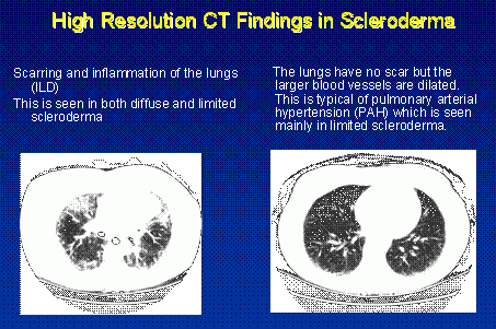
The lungs are involved in around 80% of all patients with scleroderma. Lung involvement in all its forms has emerged to be the leading cause of death and disability. Because of this fact alone, understanding the type of lung involvement and its level of activity and severity forms the central information about treatment decisions. Lung involvement occurs in both diffuse and limited scleroderma; thus all patients need to be concerned about this potential complication.
Symptoms
Involvement of the lung causes shortness of breath or fatigue during physical activity. Many patients with scleroderma become less physically active because of musculoskeletal complaints or the fatiguing nature of the illness. Hence many will not be physically active enough to actually develop shortness of breath. The doctor may inquire about your ability to do simple tasks of daily living such as climbing stairs or shopping. Other patients develop a dry, non-productive cough which also might worsen with physical activity.
In addition to the medical history, important clues about lung involvement can be obtained by a close physical examination of the heart and lungs. However, lung involvement can only be accurately assessed with appropriate use of the broad ranging battery of sophisticated laboratory testing described below.
How Does Scleroderma Affect the Lungs?
There is no single simple entity of scleroderma lung disease. Rather, it is key to understand that several different processes that are typical of the scleroderma disease process may be present and in different levels of contribution. The key elements of scleroderma lung disease involve 1) inflammation (potentially treatable); 2) lung scarring (not reversible but potentially preventable); and 3) blood vessel injury (treatable – see Pulmonary Hypertension).
ILD (Interstitial Lung Disease)
Inflammation and scarring of the lung tissue is called interstitial lung disease or ILD. The doctor may suspect ILD if the bases of the lungs make crackling sounds on stethoscope examination. ILD is best assessed by complete pulmonary function testing. These detailed tests measure lung volumes, breathing mechanics and other features of how your lungs are actually functioning. There are two key tests – the forced vital capacity (FVC) and the diffusing capacity (DLCO). The FVC measures lung volume which will be reduced if the lungs are stiffened by scar. The DLCO measures how easily oxygen makes it from the air sacs of the lung into the blood stream. The DLCO will be reduced if the air sacs are inflamed or thickened by scar AND if there is injury to the lung blood vessels.
If ILD is suspected based on history, physical examination and pulmonary function testing, the next question is determining how much inflammation (activity) is present versus how much scar (damage). The most commonly used test to determine this is a high resolution CT scan of the lungs (HRCT). This computer assisted chest X-ray can sensitively detect scarring that would be missed by simple chest X-ray and can help to assess the presence and distribution of lung inflammation.

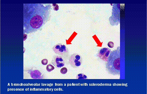
The HRCT is a valuable screening test but there are issues of both false positive and false negative results. Although not necessary in all patients, other tests to assess for the presence of lung inflammation include bronchoalveolar lavage (BAL) and/or surgical lung biopsy. BAL is done by a pulmonary specialist who introduces a slender flexible telescope into the lungs and washes up a sample of fluid from the base of the lungs. This fluid can be studied under the microscope for evidence of inflammation.

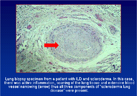
Some patients in selected situations need to take the more definitive step of lung biopsy. This is an invasive surgical procedure usually done by introducing an operating telescope through an incision in the rib cage. The tissue can be studied under the microscope and through more extensive laboratory testing to gain insight into the type of lung scarring and its activity.

Pulmonary Hypertension
In addition to scarring and inflammation, the blood vessels of the lung are frequently involved as an intrinsic part of the scleroderma disease process. The biopsy specimen above shows all three forms of lung damage active in the same patient. Pulmonary Hypertension is described in more detail in its own section. It is extremely important to consider the contribution of blood vessel damage to the individual patient’s shortness of breath since the approach to treatment is considerably different.
Natural History of ILD
The natural history of scleroderma ILD is quite variable and not all patients need treatment. In many, the ILD develops very early and very rapidly but once established can be very stable for years. In others, the ILD continues to damage the lung on an ongoing basis. This situation is critically important to recognize since prevention of lung injury becomes the linchpin issue in treatment.
Because of the inherent stability of lung function in many patients, the information about treatment is less than adequate. Many studies have been done without experimental controls and have reported stable lung function as evidence of treatment effect. This clearly overestimates the value of certain medications since many of these patients would have been stable on no treatment at all.
Closely following the pulmonary function tests is key in the decision making process. If the lung function seems stable or improved, then continued close observation is appropriate. However, if lung function is deteriorating – decisive and early treatment intervention is required.
The best studied drug is the immune system suppressant agent known as cyclophosphamide (Cytoxan®). A large controlled study done in the United States and a smaller one done in England both demonstrated that patients receiving cyclophosphamide lost lung function more slowly than patients receiving “sugar pill”. The benefit was disappointingly small but nonetheless significant. Cyclophosphamide has many important side effects including bone marrow damage, bladder irritation, infertility and cancers. The decision to use cyclophosphamide requires close attention to weighing risk versus benefit in the individual patient.
Cyclophosphamide appears effective but can be viewed as a “sledgehammer” – a strong drug with utility but lacking a specific application to the scleroderma disease process. Research moves ahead rapidly in this field seeking agents that address specific aspects of the scleroderma process.
What Can You Do to Help Yourself?
All patients benefit from simple measures that are under their own control.
- STOP SMOKING!
- Avoid passive smoke.
- Stay physically active – all patients can improve their fitness and thus preserve their function.
- Treat your esophagus correctly. Patients with esophagus problems can aspirate intestinal contents into the lung thus causing additional lung injury. See the section on Gastrointestinal Issues.
- Don’t ignore symptoms. If you feel there is a change in your breathing, inform your physician. Early diagnosis and early use of preventative treatment is the key. Lung damage, once established, cannot be reversed.
